

Amodeo D.1, De Palma I.1, Papale G.2, Nante N.2, Puccio A.2, Cevenini G.1, Messina G.2
1. Department of Medical Biotechnologies, University of Siena, Siena, Italy
2. Department of Molecular and Developmental Medicine, University of Siena, Siena, Italy
Ultraviolet C (UV-C) radiation, particularly in the peak wavelength range of 260 to 265 nm, is remarkably effective in deactivating micro-organisms. This specific wavelength range causes the formation of genetic abnormalities known as thymine dimers and uracil dimers. These abnormalities disrupt replication and transcription processes, rendering the material non-functional. Mercury UV-C lamps have a long history of effective germicidal performance. They are widely used in healthcare and water treatment applications and have a proven track record. However, conventional mercury lamps, which emit UV-C light at 254 nm, possess limitations such as size, warm-up time and inherent risks to health and the environment.
As an alternative, UV light-emitting diodes (UV-LEDs) have emerged as a viable option, offering instantaneous high-intensity light. UV-LEDs exhibit compactness, durability and economic feasibility. Furthermore, they do not contain mercury, ensuring a safer choice and maintaining a consistent output with low-temperature fluctuations. They can emit specific target wavelengths, such as the desired range of 260 to 265 nm, or very low frequencies, such as 232 nm (not too far from the 222 nm of excimer lamps), to effectively accomplish germicidal actions. Unfortunately, the more the wavelength decreases, the more the output power and efficiency of the LED falls. The main objective of this investigation was to compare the performance of four UV-C LEDs, each operating at distinct wavelength peaks, for the inactivation of three bacterial species.
Methods
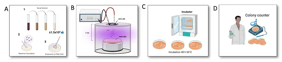
The experimental study was conducted between April and May 2023. Petri dishes, filled with generic growth medium (Plate Count Agar), were contaminated with E. coli (ATCC 8739), S. aureus (ATCC 43300) and P. aeruginosa (ATCC 27853) at a concentration of 1.5×10⁴ CFU/mL. Petri dishes then were positioned 7 cm from the LED light source, exposing them to different UV-C wavelength peaks: 232 nm, 255 nm, 265 nm and 280 nm (Figure 1). Three energy doses were tested for each microbe: 2, 4 and 5 mJ/cm2 for E. coli; 3, 4.5 and 6 mJ/cm2 for S. aureus; 3, 4.5 and 8 mJ/cm2 for P. aeruginosa.
The different energy doses for each bacterium were chosen based on data in the literature on the biocidal efficacy of UV-C radiation at 254 nm emitted by conventional mercury lamps. In particular, the authors selected three doses (for each species) to abate respectively: approx. 1 log10, 1-2 log10 and about 2 log10 of the bacterial inoculum concentration.
The statistical analysis was performed using Stata software. The mean reduction obtained by exposing the strains to the different LED wavelengths was compared using the parametric one-way ANOVA test. Results were expressed as log10 with a 95% confidence interval (CI). Statistical significance was set at 95% (p<0.05).
Results
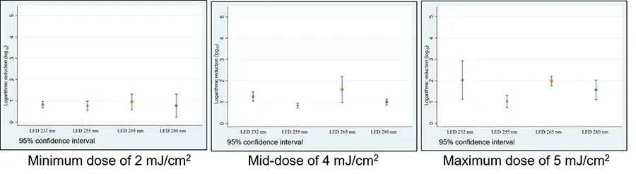
The results obtained by exposing bacteria to different wavelengths depend on both the species exposed and the dose administered. Figure 2 shows the average log10 reductions obtained in the three replicates performed on E. coli with the different LEDs at the minimum, mid and maximum doses (Figure 2). When the lowest energy dose was administered to E. coli, no significant difference was observed between the LEDs, as the reduction means all were in the same range (between 0.23 and 1.31 log10). However, when the energy dose was increased to 4 mJ/cm2, the lowest reduction was obtained for the 255 nm LED (0.84 log10; CI 95% 0.72-0.95). This was significantly different from 232 nm (1.26 log10; CI 95% 1.04-1.48) and borderline significant for 265 nm (1.60 log10; CI 95% 0.90-2.21). Even when the bacterium was exposed to the highest dose, a significant difference in inactivation level was observed between the LEDs at 255 nm (1.03 log10; 95% CI 0.74-1.32) compared to that at 265 nm (1.98 log10; 95% CI 1.76-2.21). Thus, it can be seen that as the dose administered increases, there was a greater mean bacterial inactivation common to all LEDs. However, statistical significance only was highlighted between the minimum and medium doses administered to E. coli by the 232 nm LED.
four LEDs.
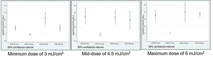
For S. aureus, a statistically significant difference in the reduction of microbial concentration was found among the 255 nm LED (0.90 log10; CI 95% 0.81-0.99) and all other LEDs, which was less effective in inactivating the microorganism regardless of the doses administered. Furthermore, at a dose of 3 mJ/cm2, the wavelengths at 255 nm and 265 nm, which caused the least and most reduction, respectively, showed significant differences from each other and with 232 nm and 280 nm LEDs (Figure 3). Similarly to E. coli, there was a clear proportionality between dose increase and mean reduction for S. aureus. Differences were statistically significant for the 232 nm and 280 nm LEDs.
four LEDs.
The results of the P. aeruginosa tests showed a significant difference in the microbial reduction values at the lowest administered dose of 3 mJ/cm2 obtained with the 265 nm LED (2.38 log10; 95% CI 2.13-2.62) compared to the 255 nm LED (1.04 log10; 95% CI 0.47-1.61) and 280 nm LED (1.69 log10; 95% CI 1.36-2.03). At the mean and maximum doses administered, the 255 nm LED again was the least effective compared to the other LEDs (Figure 4). A significant effect on bacterial inactivation with increasing dose only was observed when P. aeruginosa was exposed at 265 nm.
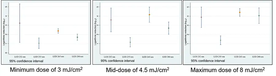
Comparing the logarithmic reduction averages obtained in the tests performed on S. aureus exposed to the highest energy dose (6 mJ/cm2) and P. aeruginosa exposed to the lowest energy dose (3 mJ/cm2), no significant differences were found between the different LED wavelengths (p<0.05).
Conclusion
The results of the study highlight the differences between the specific UV-C wavelength peaks emitted by LEDs, underlining their significant impact on the biocidal efficacy of UV light. Indeed, not all UV-C wavelengths were equally effective in inactivating the microorganisms. Even very low frequencies, such as 232 nm, seemed to be more effective than others, despite the much longer irradiation times needed to match the doses and comparable to those of LEDs emitting frequencies between 400 and 450 nm to reach the same bacterial inactivation. By identifying the key emission characteristics of LEDs, the study demonstrates the need to optimize the use of LED at different wavelengths for different applications.
The field of disinfection is undergoing a significant technological transition, marked by the move from traditional mercury-based UV-C lamps to UV-C LEDs. This transition is being driven by a number of compelling factors and offers a promising future for more efficient and environmentally friendly disinfection methods. Indeed, UV-C LEDs offer precise control over the wavelengths emitted. This flexibility allows users to tailor UV-C output to specific target areas, optimizing disinfection processes while minimizing potential damage to materials and surfaces. Furthermore, understanding the specific effects of different wavelength peaks of UV-C rays enables targeted and customized approaches to effectively neutralize bacteria, viruses and other pathogens. This knowledge paves the way for the development of advanced technologies that harness the power of specific UV-C wavelengths, optimizing their effectiveness in various biocidal applications.
References
- Bolton, J. and C. Cotton, The Ultraviolet Disinfection Handbook 2008: Amer Water Works Assn.
- Kowalski, W., Ultraviolet germicidal irradiation handbook: UVGI for air and surface disinfection. 2009: Springer.
- Boyce, J.M. and C.J. Donskey, Understanding ultraviolet light surface decontamination in hospital rooms: A primer. Infect Control Hosp Epidemiol, 2019. 40(9): p. 1030-1035.
- Sellera, F.P., et al., A systematic scoping review of ultraviolet C (UVC) light systems for SARS-CoV-2 inactivation. J Photochem Photobiol, 2021. 8: p. 100068.
- Masjoudi, M., M. Mohseni, and J.R. Bolton, Sensitivity of Bacteria, Protozoa, Viruses, and Other Microorganisms to Ultraviolet Radiation. J Res Natl Inst Stan, 2021. 126:126021.
Acknowledgements
In alphabetic order, the authors thank Bytech s.r.l., Silanna Group Pty Ltd and Stanley Electric co. Ltd for providing the LED samples used for the microbiological tests.