

Originally written by Dr. Eugene I. Gordon Ph.D., IEEE Fellow and Edison Medal Recipient, and Peter E. Gordon MSEE, in April 2012. Updated in July 2023 by Peter E. Gordon for the benefit of the UV-C and Far UV solution design and global user community. Provisional Patent has been filed.
The utility of biodosimetry in the water treatment space has been well documented since the 1970s. It has been established as the “gold standard” for determining UV system validation and a driving factor to adjust design and development of solutions to meet installation criteria for the determined operating conditions. 1 But, like other verification options, such as chemical actinometry and radiometry, it has its pros, cons and limitations. 2 A push to develop alternative approaches that will provide more accuracy with near-immediate response time that will further meet the stated objective of assuring UV dose determination and resultant efficacy in various applications 3 should be examined. One vein to mine when seeking such a capability is the commercialization of a true UV-sensitive biological indicator (BI).
A biological indicator for establishing that the sterilization process has been effective in sterilizing a given batch of surgical instruments has been the FDA-cleared holy grail for testing the efficacy of instrument sterilizers, such as autoclaves (steam) or H2O2, O3 + H2O (vapor). Endospore-based BIs particularly are appropriate for testing autoclave sterilization. 4 A good introduction to the use of BI in sterilization equipment verification is given by CDC 5 and by 3M. 6 The 3M website contains reference to an excellent review article. 7
Basically, a conventional capsule-based BI used in healthcare settings – and incidentally, also in food safety and pharma processing – involves testing for sterility a group of pathogen endospores, such as Bacillus subtilis, that have been subjected to a sterilizing procedure. Geobacillus stearothermophilus is used for autoclaves. The BI capsule is included in the group of devices being sterilized. After sterilization, the chosen test pathogens from the BI are placed in a petri dish or nutrient solution, and the extent of colony formation is determined after a period of about 48 hours and sometimes up to seven days. In a count of colonies, one colony per surviving endospore compared to the initial number of test endospores provides the sterilization rate. Sterilization is defined as a reduction in the surviving number compared to the initial number in the test by a factor exceeding 106 – the so-called −6 log10 reduction. Hence, appearance of more than a single colony in the BI test group is a failure of the sterilization process. In fact, industry practice is far more lackadaisical since the risk of imperfect sterilization is the proverbial ‘horse already out of the barn’ by the time the test results appear, and new batches of instruments have been purported sterilized in the central sterile operations of hospitals and used on patients in the operating room.
Fast process indicators invariably are used with a given batch being sterilized for convenience since they are used to only confirm that the system parameters were achieved; they do not confirm actual and uniform batch sterility. Unfortunately, as previously stated, current BI approaches do not provide real-time feedback that a batch has been sterilized. They instead are used in clinical situations to confirm periodically that the sterilization system is working properly. No current BI on the market can unassailability confirm sterility of a given batch quickly; say, in 10 minutes. If such a capability were available, reliable, consistent, accurate and reproducible – and, hence, capable of obtaining FDA clearance, it would provide a major improvement in sterilization practice globally, greatly enhancing patient safety.
It is against this background of understanding conventional and accepted uses of BIs that promoting the potential and wide utility of a UV-C and Far UV radiation-sensitive BI, rather than dose cards used for sanitation process monitoring capability, also known as a dose card 8, can be better appreciated as a more widely accepted, possibly universal, means to produce quantifiable and accurate Germicidal UV efficacy Assurance (GUVeA). Such a system could overcome documented current inefficiencies and inconsistencies in efficacy verification, at least for drinking water disinfection 9 and, by extension, also resolve gaps in standard air and surface UV sanitation verification methods. 10
Rationale for an Accurate and Rapid Read-Out BI for GUVeA
Currently, the most prolific worldwide UV-C application is for use in drinking water disinfection. With such wide adoption, there is reasonable consensus regarding certification of UV systems for drinking water treatment (USEPA, NSF 55, DVGW, ONorm for emergency management, etc.), and there are some established installation validation techniques. However, there are very few, if any, regulations governing other uses for UV-C and FarUV implementation and none for demonstrating assurance of UV systems for treating the built environments’ air and surfaces. A separate article, perhaps part 3 in this series, will describe how the technology to be discussed in part 1 and part 2, also could be deployed in water disinfection systems to supplant reliance on the aforementioned indirect means. For now, part 1 and part 2 of the series will focus on the reality that even with the advent of the recently released ASHRAE 241 standard, which only in passing mentions the capabilities of UV-C air treatment to substitute for room air ventilation, there remains a wide chasm in universally accepted performance metrics and prove-in guidance, resulting in a problematic credibility gap. 11 One notable exception is a plethora of studies demonstrating the lowering of infection rates with treatment in hospitals due to specific infectious pathogens utilizing post-cleaning treatment of surfaces with mobile UV-C radiation-emitting platforms. 12 These studies were conducted without the benefit of a universal quality assurance tool, yet still produced statistically acceptable conclusions. Such a tool would enable the production and verification of efficacy claims under varying operational conditions.
In what follows, an automatable technical approach is described for such a BI for GUVeA that gives the equivalent feedback of a petri dish colony count and potential failure to meet minimum acceptable levels indication in less than 10 minutes after exposure. The validity of the science and its ability to replicate success criteria, which could be that the procedure becomes defined as detecting no or minimal equivalent colony count, does not rely on counting bacterial colonies produced by endospores that were not inactivated during an imperfect disinfection process. Rather, validity is shown by detecting the locked-up dipicolinic acid (DPA) released by germinating endospores that were not inactivated during the process. This is a well-understood phenomenon, with the earliest paper found on this subject published in 1978 by Scott et al. 13 The authors describe counting endospores that survive the imperfect sterilization process and that begin the activation process by germinating when exposed to nutrient. The number of endospores that germinate is strictly related to the number that survive, but the authors do not relate the number that germinate to the number that can reproduce and produce colonies. The Scott device can be described as useful as a germination assay, but it still relies on future manual counting.
A key next step would be to demonstrate that the germination rates and colony forming rates are strictly related, so measuring the endospore germination rate is equivalent to counting colonies once the relationship is understood. This trajectory then would lead to a unique capability for realizing a functional GUVeA, satisfying the user base that UV-C and Far UV disinfection to the degree that is necessary to provide adequate safety has been verified throughout solution operation. 14
Photoluminescent Staining
A thorough overview now provides the understanding to appreciate how such a GUVeA would bio-photonically function. It is well known that applying a nutrient such as L-alanine (or alternately, I. asparagine or glucose) to a viable, hibernating endospore triggers the start of the activation process that results in the conversion of the endospore to a vegetated bacterium. The first step in activation – germination – is known to i cause the complete expulsion of dipicolinic acid (DPA) 15, which is Pyridine-2, 6-dicarboxylic acid, from the nucleus of the endospore into the external nutrient surrounding the endospore. 16 The DPA is an important and necessary constituent of the nucleus material of a hibernating endospore but is absent from the vegetated form of the bacterium. Indeed, it can be said loosely that the DPA in the nucleus is what keeps the endospore from activating. On the other hand, nutrient triggers the conversion of the endospore to a vegetated bacterium. Once the endospore detects a suitable environment for survival of the vegetated form of the bacterium, it begins the process of activating and converting to the vegetated form by expelling the DPA from the nucleus into the region around the endospore and germinating.
As will be seen, this characteristic offers some important options for quickly detecting germinating endospores in a group that supposedly has been permanently inactivated by exposure to UV-C or other low-temperature systems. For example, Panigrahi 16 shows that the DPA as a ligand (cell adhesion molecule) in conjunction with Tb3+, Eu3+ and Dy3+ in the nutrient exhibits green photoluminescence under illumination by UV. Specifically, the DPA molecule absorbs an incident UV photon, is excited and non-radiatively transfers the excitation energy to a ground state Tb3+ atom within a Tb3+ lanthanide molecule to which the DPA molecule has adhered. The Tb3+ atom is excited to a radiating excited state. Within the millisecond following excitation, the Tb3+ radiates the absorbed energy into a narrow band of green light, which readily is detected. Photoluminescence from Tb-DPA is well known. For the case at hand, this does not occur to any significant extent without the presence of DPA outside the endospore. Hence, the appearance of the green photoluminescence signals the appearance of expelled DPA outside the endospore and the start of the germination. It is an indicator that viable endospores are starting to activate by virtue of detecting a favorable environment for survival of the vegetated form of the bacterium. An important paper on work at the California Institute of Technology, Jet Propulsion Laboratory 17, and based on the earlier work of Panigrahi advancing the method, describes a technique for rapid determination of the extent of failure of endospore inactivation following sterilization by measuring the cumulative, visible (green) photoluminescence from the surviving, nominally undamaged endospores of Bacillus Strophies and Bacillus Subtilis following a sterilization. The observation is made during the process of germination in a nutrient, terbium3+/L-alanine agarose. 18
In summary, associated with initiation of germination, the DPA leaves the nucleus of the nominally undamaged endospore and passes into the nutrient region around the endospore. This region contains Tb3+, in the form of a lanthanide – Tb (DO2A)+, and the DPA combines with the Tb3+ lanthanide to form Tb-DPA complexes. The DPA molecule will absorb energy from incident UV illumination; none radiatively transfer the excitation energy to the attached Tb3+ ion, which then emits photons of narrow-band green light.
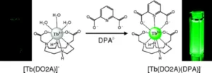
Figure 1 illustrates two cases associated with UV illumination of the nutrient:
- No green light and, therefore, no DPA present – implying no viable endospores, even in the presence of nutrient
- Green light and, therefore, DPA present – implying viable endospores responding to the presence of nutrient
This is done at 37° C, and it takes 10 minutes to observe the full count of green light-emitting, germinating endospores. This also corresponds to the time when germination is complete. It does not require division of the newly vegetated bacteria. Hence, the process relies only on endospore germination, a 10-minute process, and often will not proceed to fully vegetated bacteria to allow detection and evaluation of the actual count of surviving, fully viable endospores. It is known that some endospores, in the presence of nutrients, will start to activate but not complete the full activation process to become viable vegetated bacteria. They have been damaged, but not fully inactivated, and will start to germinate.
Germination is induced in the BI by transferring the nominally sterilized endospores onto a quartz slide with a sample well containing the agarose nutrient plus Tb3+. Photoluminescence (from individual green spots corresponding to germinating endospores) is induced in the Tb-DPA around the endospore by illumination of the slide with UV from a pulsed UV source. The green photoluminescence is imaged to determine the number of endospores that germinated, as described in the patent reference of Footnote 13. The number germinating can be counted, for example, with a microscope fitted with an electronic camera by photographing the emitted light as described in the paper of Footnote 17. Hence, it can be a fully automated, fast technique, taking not more than 10 minutes. US patent 7,306,930, filed in 2002 and assigned to CIT, covers this technique. 19
Detection Method Using a Pulsed UV-C LED Sub-System
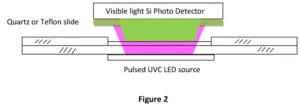 Alternately, as illustrated in Figure 2, a lanthanide stain such as Tb3+ that combines with DPA and reflects or absorbs in a narrow band to color the germinating endospore can be used. Under rapid-pulse UV illumination (e.g., from UV LEDs pulsed at approximately 500 pps), the collective group of endospores would emit pulsed, green light if it contains one or more germinating endospores because the lifetime of the photoluminescence is about one millisecond. The pulsed green light would be collected by a large-area, narrow-band, green-filtered photo detector (silicon) or a photomultiplier, and the photo current would be homodyne-detected and integrated to yield an output signal proportional to the intensity of the collected green light output with minimal noise. Hence, the pulsed green light output can be detected with extremely high sensitivity. With good design, the signal would indicate how many germinating live endospores are on the slide, including as few as none.
The process can include placing the test coupon after sterilization directly into the well of the quartz slide and then dropping the nutrient solution or nutrient plus Tb3+ into the well to initiate the germination and to provide for the photo-luminescence detection.
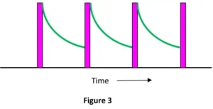 In this case, a pulsed UV source, possibly a UV-emitting, large area LED, is placed directly under the well and transmits through the quartz substrate to illuminate the sterilized sample in the bottom of the well. In this example, the configuration is fixed and the special quartz slide with the test endospore is removed from the BI after exposure and placed in a slot above the LED illumination source and below the photo detector. The LED is pulsed on and off in a short pulse at a rate of 500 to 1,000 times per second, and the pulsed UV-C illuminates the endospores and the thin layer of nutrient solution. The photoluminescence from a single germinating endospore surrounded by DPA originates after a brief formation delay from a huge number of Tb3+-DPA molecules surrounding the germinating endospore. Since the lifetime of the radiating transition is about one millisecond, the narrow-band green radiation from a single endospore and a single UV-C pulse decays exponentially, as shown in Figure 3. The green radiation is incident on the photo detector covered by a green filter, so essentially the only radiation entering and detected by the photo detector is the pulsed, periodically varying, green photoluminescence. The output oscillatory photo detector current is homodyne-detected to eliminate noise and extraneous background signals and integrated to produce a DC signal proportional to the integration time and the number of germinating endospores. Use of a photomultiplier enhances sensitivity.
The system, particularly once FDA 510K pre-market cleared as a true GUVeA (which will be further discussed in Part 2), can be calibrated to establish the exact number of germinating endospores. This would be a leap in germicidal accuracy, beyond that provided by a dose monitors deployed today 20 and provide efficacy assertions supporting ongoing efforts by industry to build confidence among a user base seeking expanded guidance and oversight by regulatory agencies. 21
Dr. Eugene I. Gordon Ph.D, IEEE Fellow and Edison Metal Recipient, was co-inventor of the Argon Ion laser (applied to treating diabetic retinopathy), the CCD Imaging device (enabling fax machines and night vision googles), and Video Conferencing (The PictuePhone was first used during the Moon Landings).
Peter E. Gordon is director of business development, CudoForm Tech, Senko Advanced Components, Inc. and is primarily focused on Photonic IC Optical I/O to Fiber Optic connectivity solutions for next-generation Ethernet Switches in Data Centers and AI chip-to-chip, node-to-node in high-speed computers. He is a member of EPIC, Optica, the IEEE, ASHRAE, the UL8802 standards group and the IUVA, and an elected member of the Board of Education in Weston, Connecticut. For more information, email peter.gordon@senko.com for Senko Advanced Components, Inc. or pgordon051163@gmail.com for LiTeProducts LLC.
References
- Malley et. al., Inactivation of Pathogens with Innovative UV Technologies, American Water Works Association, 2004 and private conversation with Professor Malley regarding the rationale for alternative approaches.
- Uppinakudru et. al, Critical assessment of optical sensor parameters for the measurement of ultraviolet LED lamps, Measurement Volume 196, 15 June 2022
- https://www.iuva.org/2023-GUV-WS
- http://en.wikipedia.org/wiki/Endospore This is a short, excellent background article on endospores, which are the chosen pathogen for testing efficacy of steam sterilization. Steam is particularly effective in inactivating endospores. Endospores are typically used in BIs.
- A good review of BI practice is given by CDC. http://www.cdc.gov/hicpac/disinfection_sterilization/13_11sterilizingpractices.html
- http://solutions.3m.com/wps/portal/3M/en_US/EveryLoadMonitoring/Home/ELM/ScienceBI/?MDR=true. The website allows downloading a comprehensive summary of the technology.
- Sailaja Chandrapati, and MarthaYoung, To Kill or Not to Kill, MANAGING INFECTION CONTROL, November 2008, http://multimedia.3m.com/mws/
- Masse et al., Comparing and optimizing ultraviolet germicidal irradiation systems use for patient room terminal disinfection: an exploratory study using radiometry and commercial test cards, Antimicrobial Resistance and Infection Control (2018) 7:29
- Wright, H., M. Heath, T. Brooks, AND Jeff Adams. Innovative Approaches for Validation of Ultraviolet Disinfection Reactors for Drinking Water Systems. U.S. Environmental Protection Agency, Washington, DC, EPA/600/R-20/094, 2020
- www.nist.gov/system/files/documents/2020/03/23/Panel%20III%20Matthew%20Hardwick%20presentation.pdf
- https://www.ashrae.org/about/news/2023/ashrae-approves-groundbreaking-standard-to-reduce-the-risk-of-disease-transmission-in-indoor-spaces
- https://www.uvdi.com/hospital-studies/ and https://sy-g.com/wp-content/uploads/2020/07/Xenex-Effectiveness-Summary-Studies.pdf provide examples.
- I. R. Scott, D. J. Ellar, Study of calcium dipicolinate release during bacterial spore germination by using a new, sensitive assay for dipicolinate, J. Bacteriol 135: 1978, 133-137, ABSTRACT, The release of calcium and dipicolinic acid from spores of Bacillus megaterium KM during L-alanine-induced triggering of germination has been studied using a new, simple, and rapid assay for dipicolinic acid capable of detecting a concentration of 0.5 micron. The release of both calcium and dipicolinate started within seconds of exposure of the spores to L-alanine, thus preceding other measurable changes associated with germination. From the earliest times, the two substances were released in equimolar quantities, although later in germination calcium predominated.
- Eadie et al., Far- UV-C (222 nm) efficiently inactivates an airborne pathogen in a room-sized chamber, Scientific Reports volume 12, Article number: 4373 (2022)
- Up to 15% of the dry weight of the endospore consists of calcium dipicolinate within the core. DPA is an important ingredient of the endospore. http://aem.asm.org/cgi/content/abstract/67/3/1274
- B. S. Panigrahi, A fluorimetric study of terbium, europium and dysprosium in aqueous solution using pyridine carboxylic acids as ligands, Journal of Alloys and Compounds, Volume 334, Issues 1-2, 28 February 2002, Pages 228-23
- Pun To Yung and Adrian Ponce, Fast Sterility Assessment by Germinable-Endospore Biodosimetry, Applied and Environmental Microbiology, Vol. 74, No. 24, Dec. 2008, p. 7669-7674
- ML Cable, JP Kirby, K Sorasaenee, HB Gray, and A. Ponce, Bacterial spore detection by [Tb3+ (macrocycle) (dipicolinate) luminescence. J. Am. Chem. Soc. 129:2007, 1474-1475 Abstract Dipicolinic acid (DPA) is a unique constituent of bacterial spores, a dormant form of Bacillus and Clostridium, which can be detected using DPA-triggered Tb3+ luminescence. [Tb(DO2A)]+ improves the sensitivity of bacterial spore detection over Tb3+(aq) owing to the exclusion of coordinated water molecules and represents the first step toward construction of a DPA receptor site with enhanced binding selectivity. The title ternary [Tb(DO2A)(DPA)]- complex was structurally characterized and features two DO2A-DPA interligand hydrogen interactions that stabilize the complex. http://pubs.acs.org/toc/jacsat/129/6 A patent #5,876,960 dated March 2, 1999, David L Rosen; Bacterial spore detection and quantification methods predates the Cable et al patent but misses the point that DPA does not leave the endospore unless nutrient is present. In addition, it never claims nutrient.
- US Patent 7306930, issued Dec 11, 2007, was filed in 2002, assigned to CIT, Ponce, Adrian, Venkateswaran, Kasthuri, J.,Kirby, James Patrick, ‘Method bacterial endospore quantification using lanthanide dipicolinate luminescence’
- Nardell et at., Dynamic Breathing Zone Dose Monitoring of Room Occupants: A New Approach to Demonstrating Safe and Effective Germicidal UV (GUV) Air Disinfection, A51 MANAGEMENT AND IMPACT OF THE COVID-19 PANDEMIC ON RESPIRATORY HEALTH
- https://finance.yahoo.com/news/xenex-makes-novo-petition-fda